In This Article
- The Discovery That Is Rewriting Textbooks
- What We Thought We Knew — and Why We Were Wrong
- Why Do Brain Cell Axons Look Like Pearls, Not Tubes?
- What This Means for Brain Disease Research
- The Big Questions Scientists Still Need to Answer
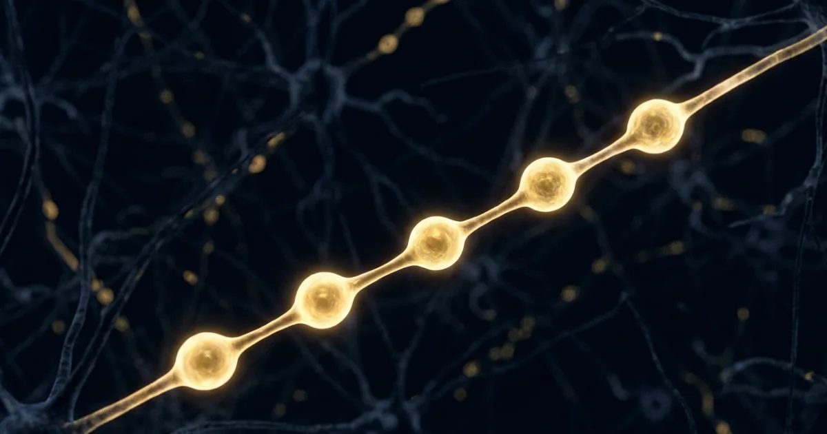
Every student who has ever opened a biology textbook has seen the same picture: a brain cell with long, smooth, tube-like arms stretching out to connect with other cells. That image has been the accepted truth for over 100 years. Scientists at Johns Hopkins University just proved it is wrong. The arms of brain cells — called axons — are not smooth tubes at all. They look more like a string of pearls. And that single correction could change how we study the brain, and how doctors one day treat diseases like Parkinson's.
The Discovery That Is Rewriting Textbooks
Axons are the long, thin extensions of brain cells that carry electrical signals from one neuron to another. Think of them as the cables of the brain — connecting everything together, allowing you to think, remember, move, and feel. For over a century, scientists assumed these cables were smooth cylinders. That assumption made it into every textbook, every diagram, every classroom poster. Nobody really questioned it, partly because axons are almost impossibly small — about 100 times thinner than a single strand of human hair. Seeing them properly required technology that simply did not exist.
What We Thought We Knew — and Why We Were Wrong
Standard electron microscopy — the technique scientists used for decades — works by firing beams of electrons at a cell to map its shape. The problem is that preparing a cell for this process involves dehydrating it first, which shrivels and distorts the structure. One of the lead researchers on this study, Shigeki Watanabe, described it simply: dehydrating neurons for imaging is like drying a grape into a raisin. The raisin does not look like the grape. Scientists thought they were studying the true shape of axons. They were actually studying a version that had been warped by the preparation process. That is a very important difference. And it took a smarter method to finally catch the mistake.
Why Do Brain Cell Axons Look Like Pearls, Not Tubes?
The Watanabe lab at Johns Hopkins School of Medicine used a technique called high-pressure freezing electron microscopy. Instead of dehydrating neurons, they froze them — locking the cells in their natural, living state before imaging them. The results were a surprise. Across tens of thousands of images of three different types of mouse neurons — lab-grown cells, cells from adult mice, and cells from mouse embryos — the same shape appeared every time. The axons were not smooth. They had repeating, bubble-like swellings along their length. The team named these bulges "nonsynaptic varicosities." A follow-up study published in Biophysical Journal in 2025 confirmed the same pearl-like structure in multiple other neuron types — including human cortical neurons. This is not a mouse quirk. This is how our own brain cells are built.
"Axons are the cables that connect our brain tissue, enabling learning, memory, and other functions."
— Shigeki Watanabe, Associate Professor · Johns Hopkins University School of Medicine · Nature Neuroscience, 2024What This Means for Brain Disease Research
Here is what makes this truly interesting. For years, doctors and researchers noticed bead-like swellings on axons in patients with diseases like Parkinson's disease, Alzheimer's, and other neurodegenerative conditions. The assumption was always that these swellings were a sign of damage or disease — something abnormal happening to an otherwise smooth cable. This new research turns that thinking on its head. If healthy axons are already pearl-shaped by nature, then researchers need to completely rethink what "abnormal" actually looks like. The swellings seen in diseased brains may differ in a very specific way from healthy ones, rather than simply existing where they should not. For India, where neurological diseases like Parkinson's affect millions — and where research into early detection is growing fast — this kind of foundational reset in understanding has direct implications for future diagnostics and drug development.
The Big Questions Scientists Still Need to Answer
This discovery is not the final word — it is the beginning of a new set of questions. The studies so far have focused primarily on nonmyelinated axons, which are neurons without the insulating myelin sheath. Myelinated axons — which make up a large portion of the nervous system — still need to be examined with the same freezing technique. A separate 2025 study published in Neuron extended these findings to living brain tissue from epilepsy surgeries, which is encouraging. But the full picture across the human nervous system is still being built. Scientists also want to understand exactly how the pearl shape affects the way electrical signals travel and how these structures change during disease. One thing is already clear, though: a lot of what was written in textbooks about the basic shape of brain cells is going to need to be rewritten.
- Pearls, not tubes — Brain cell axons have a natural pearl-like shape, confirmed across multiple neuron types in mice and humans, overturning over a century of assumption.
- Old method, wrong picture — Traditional dehydration-based electron microscopy distorted axon shape during preparation; freezing techniques have now revealed the true structure.
- Disease research changes — Swellings on axons in diseases like Parkinson's must now be understood differently, since healthy axons already have bumps — making the baseline itself brand new.
"Understanding the structure of axons is important for understanding brain cell signalling. Axons are the cables that connect our brain tissue, enabling learning, memory, and other functions." — Shigeki Watanabe, Nature Neuroscience, 2024.
📄 Source & Citation
Primary Source: Mougkogiannis P, Watanabe S, et al. (2024). Nonsynaptic varicosities reveal the true morphology of mammalian axons. Nature Neuroscience, 27, 2325–2338. https://doi.org/10.1038/s41593-024-01813-1
Authors & Affiliations: Shigeki Watanabe (Johns Hopkins University School of Medicine, Department of Cell Biology and Neuroscience) and collaborators from Johns Hopkins and international partner institutions.
Additional Study: Follow-up findings in nonsynaptic varicosity structure published in Biophysical Journal (2025) and extended to living human tissue in Neuron (2025) via zap-and-freeze electron microscopy of epilepsy surgery samples.
Data & Code: Supplementary imaging datasets available via the journal's online portal at Nature Neuroscience and Johns Hopkins institutional repository.
Key Themes: Neuron Morphology · Axon Structure · Brain Disease · Electron Microscopy · Textbook Revision
Supporting References:
[1] Watanabe S et al. (2020). Flash-and-freeze: a novel technique to capture the membrane dynamics associated with synaptic vesicle fusion. Frontiers in Synaptic Neuroscience, 12:24.
[2] Lapicque L. (1907). Recherches quantitatives sur l'excitation électrique des nerfs traitée comme une polarisation. Journal de Physiologie et de Pathologie Générale, 9:620–635. [The foundational 1907 integrate-and-fire neuron model that informed decades of axon theory.]
[3] Bhatt DL et al. (2009). Axonal swellings and degeneration in mice overexpressing the metabotropic glutamate receptor 1. Brain, 132(Pt 1):258–269. [Prior research linking axon swellings to neurodegeneration — context now reframed by this discovery.]
No comments yet. Be the first to share your thoughts.
Leave a Comment