In This Article
- The Brain Signal That Arrives Before the Decision
- Why Studying Social Behavior in a Moving Brain Is So Hard
- How Did Scientists Read a Fish's Mind During a Real Friendship?
- What the Neural Patterns Actually Look Like
- The Questions That Still Need Answering
Something in your brain decides you want to walk over to a friend — before your legs start moving and, it turns out, before you are even fully aware of it. A new study published in Nature Communications has caught this exact moment in a living brain, watching thousands of neurons shift into a distinct pattern several seconds before a fish moves toward a companion. The research pinpoints a small cluster of brain cells, called the pallium, as the region that starts the whole process — and shows that without it, the drive to approach others simply vanishes.
The Brain Signal That Arrives Before the Decision
Think of the moment just before you cross a room to greet someone. You feel a pull, a quiet decision forms, and then your body moves. For a long time, scientists assumed the key brain activity happened right when movement began, or just after. This study challenges that idea directly.
Researchers at The Hebrew University of Jerusalem's Edmond and Lily Safra Center for Brain Sciences found that social approach behavior — the act of physically moving toward another individual — is predicted by a very specific and coordinated pattern of brain activity. That pattern appears several seconds before any movement happens. It is not random background noise. It is a reliable, repeatable neural signature.
Why Studying Social Behavior in a Moving Brain Is So Hard
Here is the challenge scientists face: the best tool for watching the brain in fine detail requires the animal to hold very still. But social behavior, by definition, involves movement and interaction. These two requirements seem to pull in opposite directions.
In mammals like mice or monkeys, there is no technology yet that can record millions of individual brain cells at once during live social interaction. The brain is too large, too dense, and too deep inside the skull. This is why so much of what we know about social neuroscience comes from either simplified artificial scenarios or from studying brain activity only after behavior has already happened.
How Did Scientists Read a Fish's Mind During a Real Friendship?
The team solved the stillness-versus-movement problem with an elegant setup. One fish was gently held in place at the center of a small water-filled plate using a technique that kept its head fixed but left its tail completely free to move. A second fish swam freely in a surrounding ring of water, visible to the first fish across a transparent barrier.
The held fish could see its companion moving naturally. It could not smell it or feel it — only watch it. This gave the scientists precise control over what information the brain was actually receiving. Meanwhile, a laser-based microscope called a two-photon microscope scanned through the brain of the held fish in layers, recording the activity of every visible neuron in real time.
The result was a continuous, live-action movie of more than 12,000 brain cells firing as the fish watched its companion swim around. Every time the held fish twitched its tail to "turn" toward its companion, the researchers could compare exactly what the brain had been doing in the seconds leading up to that moment.
"These distributed patterns enabled accurate decoding of upcoming social actions within each region, and accounted for inter-individual variability in approach behavior."
— Lifshitz et al., The Hebrew University of Jerusalem · Nature Communications, 2026What the Neural Patterns Actually Look Like
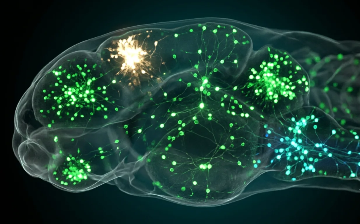
The most striking finding was the shape of the signal. Before the fish moved toward its companion, a small group of cells in the pallium — a region of the outer brain thought to be similar to the human hippocampus and amygdala — became more active. At the same time, cells in the midbrain and hindbrain, the lower and more ancient parts of the brain, became quieter.
This coordinated pattern, one part of the brain turning up while another turns down, was specific to social approach. When the fish moved away from its companion, or when scientists replaced the real fish with a moving dot following the same path, the pattern disappeared entirely. The brain knew the difference between a real companion and a visual imitation.
To confirm the pallium was not just a bystander, the team used a laser to precisely destroy a small number of pallial neurons — an average of 46 cells per fish. After 24 hours of recovery, the fish showed a measurable drop in social interest. More tellingly, the predictive brain pattern in the midbrain and hindbrain also disappeared. Remove the pallium's contribution and the whole distributed network goes quiet. [INTERNAL LINK: how the amygdala processes social cues]
The Questions That Still Need Answering
The study has important limits that the authors are clear about. The pallial neurons were identified and removed based on their physical location, not on confirmed knowledge of their function or connections. The molecular identity of these cells, what they release, which other neurons they talk to, remains unknown. It is possible that nearby, unaffected neurons were partly compensating for the loss.
The experiment also used fish that were about two weeks old, an age when social behavior is still developing. Whether the same patterns hold in adults, or whether experience and age sharpen or change the signal, has not yet been tested. The researchers also note that about a third of fish in their study showed very little social behavior at all, which suggests this brain system varies considerably between individuals even within a single species.
What comes next is the harder question: do these principles hold in a mammalian brain? Can we find the same type of anticipatory, distributed social signal in a mouse, a monkey, or a person? The zebrafish approach cannot simply be copied in those animals, but it points researchers toward what to look for.
- The pallium drives the network — Removing a tiny number of pallial cells wiped out the predictive social signal across the entire brain, not just locally.
- The signal is socially specific — The same brain pattern did not appear when a moving dot replaced the real companion, confirming this is not just a general movement signal.
- Individual differences are visible in the brain — Fish that showed stronger neural distinction before approach movements were more social overall, suggesting this pattern underlies personality-level variation.
"Our findings uncover a distributed yet coordinated neural mechanism underlying social interaction." — Lifshitz, Prag, Livneh, Moshkovitz, Karmi, and Avitan, Nature Communications, 2026.
📄 Source & Citation
Primary Source: Lifshitz I, Prag A, Livneh N, Moshkovitz M, Karmi A, Avitan L. (2026). Distinct distributed neural dynamics predict pallium-dependent social approach. Nature Communications, 17, 4848. https://doi.org/10.1038/s41467-026-71666-8
Authors & Affiliations: Imri Lifshitz, Asia Prag, Netta Livneh, Maayan Moshkovitz, Abeer Karmi, and Lilach Avitan (Edmond and Lily Safra Center for Brain Sciences, The Hebrew University of Jerusalem, Israel)
Data & Code: All source data and analysis code are available at Mendeley Data
Key Themes: Social neuroscience · Zebrafish brain imaging · Pallium · Distributed neural networks · Approach behavior
Supporting References:
[1] Harpaz R et al. (2021). Precise visuomotor transformations underlying collective behavior in larval zebrafish. Nature Communications, 12, 1–14.
[2] Zada D et al. (2024). Development of neural circuits for social motion perception in schooling fish. Current Biology, 34, 3380–3391.
[3] Dreosti E et al. (2015). Development of social behavior in young zebrafish. Frontiers in Neural Circuits, 9, 39.
No comments yet. Be the first to share your thoughts.
Leave a Comment